Home-Scientists Grow Functional Human Gut Tissue With Its Own Nervous System
Scientists atCincinnati Children’s Hospital Medical Centerhave developed a new method for growing large-scale human gastrointestinal tissue in the laboratory, one that spontaneously generates its own functional nervous system without requiring the complex, multi-step assembly processes that have long slowed progress in the field. Published inNature Biomedical Engineering, the work represents a key step toward transplantable gut tissue for patients with intestinal failure and related conditions.
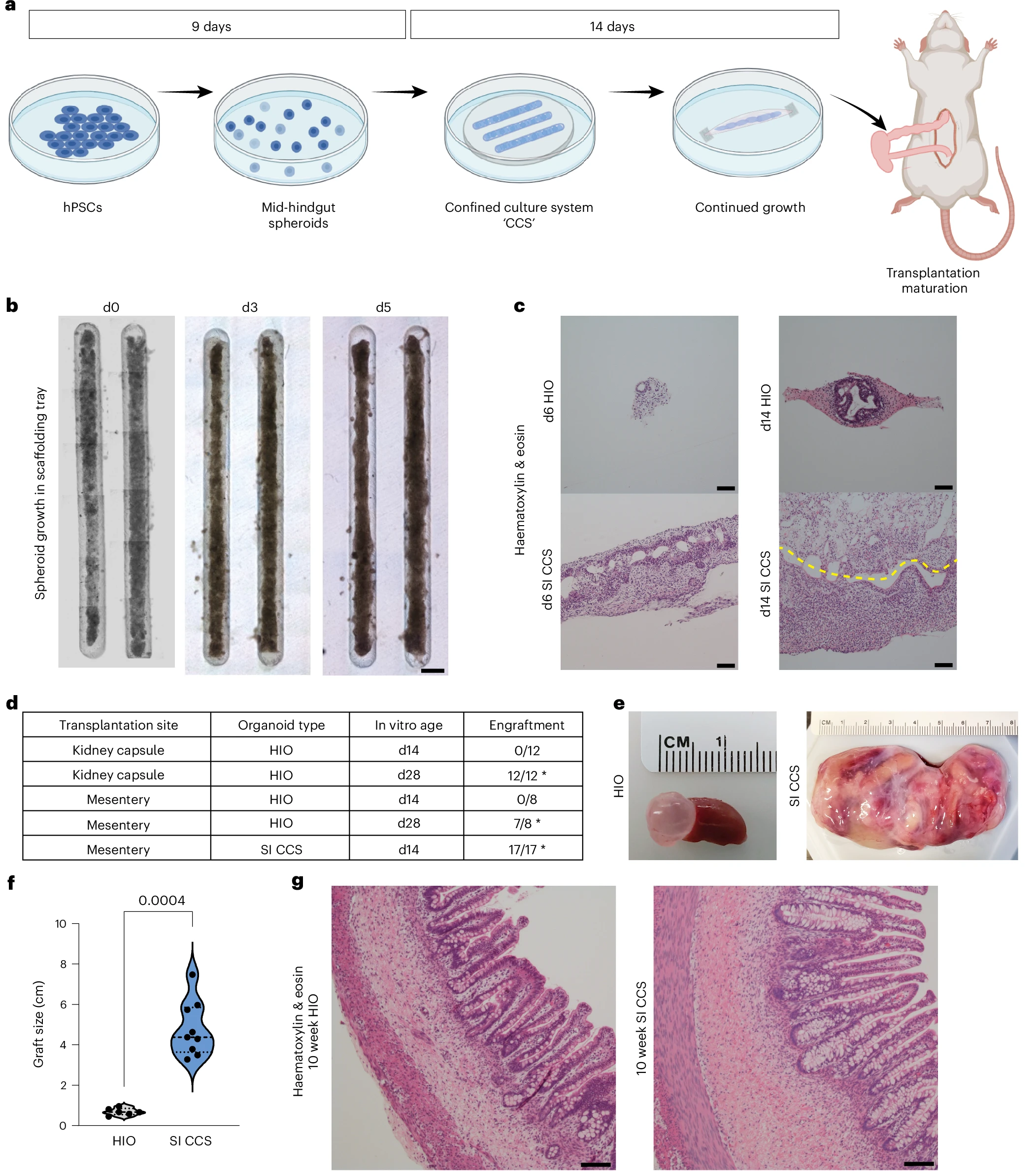
The approach, called the confined culture system (CCS), uses a 3D printed scaffolding tray to guide the fusion and growth of thousands of stem cell-derived spheroids into elongated intestinal, colonic, or gastric structures.
Rather than growing organoids one by one, the standard method, which typically produces tissue roughly one millimeter across after four weeks, the CCS loads approximately 4,000 spheroids into defined lanes, prompting them to merge into a unified structure within days. After ten weeks of transplantation into immunocompromised rats, the resulting tissues reached widths of up to eight centimeters, roughly ten times larger than those produced by conventional protocols.
A Nervous System That Builds Itself
CCS tissues developed an enteric nervous system, the network of neurons governing gut motility, fluid regulation, and other digestive functions, without externally added neural cells. Previous methods required separately differentiated neural crest cells to be combined with intestinal spheroids, a process known as an assembloid approach.
In CCS tissues, both excitatory and inhibitory neuron subtypes emerged spontaneously, confirmed through protein expression analysis, single-cell RNA sequencing, and electrophysiological testing.
Organ bath assays showed that the CCS tissues produced rhythmic contractile activity comparable to adult human intestinal samples, and responded robustly to nerve stimulation, responses that were significantly reduced when neuronal activity was blocked with tetrodotoxin, confirming the ENS was driving the contractions.
Earlier assembloid tissues showed minimal response to the same stimulation, suggesting that the CCS approach produces a more functionally mature nervous system than methods that assemble ENS components separately.
From Bench to Body: Integration With Host Tissue
Source: 3D Printing Industry
